Civil Rights
Movements, leaders, victories and the continuing fight for equality.

Explore the people, places, events, achievements, struggles and stories that shaped our journey.
Movements, leaders, victories and the continuing fight for equality.
Innovation, patents, science, technology and world-changing contributions.
Pioneers, champions, Negro Leagues, records, activism and excellence.
Meet the people whose lives, choices and achievements shaped the journey.
Black towns, communities, institutions and places where history happened.
Moments that changed communities, movements, institutions and the nation.
In 1968 Shirley Chisholm became the first Black woman elected to the United States Congress. In 1972 she launched a campaign for the Democratic presidential nomination, breaking another political barrier.
MORE →Reflects the personal views, recollections, and perspective of the author, Mike Davis.
This is a personal recollection on the Move fire on May 13, 1985
 | This article includes a list of general references but lacks sufficient corresponding inline citations. (March 2011) |
| Osteoid | |
|---|---|
 | |
| Identifiers | |
| FMA | 66830 |
| Anatomical terminology | |
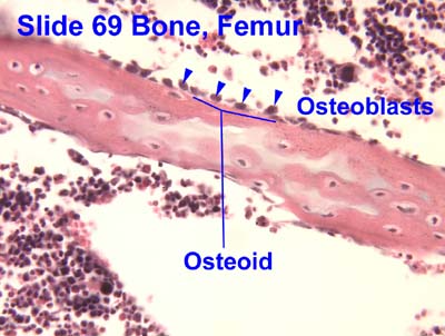
In histology, osteoid is the unmineralized, organic portion of the bone matrix that forms prior to the maturation of bone tissue.[1] Osteoblasts begin the process of forming bone tissue by secreting the osteoid as several specific proteins. The osteoid and its adjacent bone cells have developed into new bone tissue when it becomes mineralized.[citation needed]
Osteoid makes up about fifty percent of bone volume and forty percent of bone weight. It is composed of fibers and ground substance. The predominant type of fiber is type I collagen and comprises ninety percent of the osteoid. The ground substance is mostly made up of chondroitin sulfate and osteocalcin.[citation needed] Osteoblasts synthesize and secrete osteoid as an unmineralized organic matrix, and when the osteoid becomes mineralized through deposition of calcium salts, it transforms into mature bone tissue.[2]
Osteoid is primarily composed of Type I collagen and ground substance.
Type I collagen comprises approximately 85-95% of the organic matrix,[3] providing the structural scaffold and tensile strength necessary for bone formation.[4] These collagen fibers form a dense, highly cross-linked network that serves as the foundation for subsequent mineralization.[5]
The collagen molecules are arranged in layers that alternate parallel and orthogonal to the axis of stress loading, creating a sophisticated composite structure.[5]
The ground substance of osteoid comprises approximately 10% of the bone matrix[6] and includes non-collagenous proteins:
Osteoid is synthesized and secreted by osteoblasts, specialized bone-forming cells.[7] Osteoblasts are large cuboidal cells characterized by abundant rough endoplasmic reticulum, reflecting their high synthetic activity.[4]
At their apical surface, osteoblasts secrete large amounts of type I collagen and smaller amounts of matrix organizing proteins, including osteocalcin and osteopontin.[5] The newly secreted osteoid forms a hydrated protein matrix layer between the mineralization front and the osteoblast layer.[5][7]
The synthesis of bone matrix occurs in two main steps: deposition of organic matrix (osteoid) and its subsequent mineralization.[7] During active bone formation, osteoblasts continuously produce osteoid matrix until mineralization occurs.
The transformation of osteoid into mineralized bone involves the deposition of hydroxyapatite crystals [Ca10(PO4)6OH2] within the organic matrix.[6]
Mineralization occurs in two phases: the vesicular phase and the fibrillar phase.[7]
Vesicular phase: Matrix vesicles, measuring 30-200 nm in diameter, are released from the apical membrane of osteoblasts into the newly formed bone matrix.[7][3] These vesicles contain alkaline phosphatase, adenosine triphosphatase (ATPase), and inorganic pyrophosphatase, and act as seeding sites for hydroxyapatite crystal formation through localized enzymatic accumulation of calcium and phosphate.[3]
Role of alkaline phosphatase: Osteoblasts secrete alkaline phosphatase, which participates in bone mineralization by hydrolyzing pyrophosphate, an inhibitor of mineralization, thereby increasing local inorganic phosphate availability for hydroxyapatite formation.[2][4]
Crystal propagation: Crystal growth proceeds from initial foci in matrix vesicles to form spheroids, which gradually coalesce to form a network of apatite crystals.[3] As the matrix matures, hydroxyapatite microcrystals are organized into a sophisticated composite within the collagen layer by nucleation in the protein lattice.[5]
Defective mineralization of osteoid leads to several important clinical conditions characterized by accumulation of unmineralized matrix.
Rickets: Occurs in children when defective mineralization affects the growth plate and osteoid.[8] The condition results from calcium or phosphate deficiency, either isolated or secondary to vitamin D deficiency.[9] In rickets, the mineralization defect leads to accumulation of osteoid in bone tissue below the growth plate, resulting in weak bones and deformities.[9]
Osteomalacia: The adult equivalent of rickets, osteomalacia is characterized by softened bones due to impaired mineralization of osteoid.[10] Vitamin D deficiency is the most common cause, though calcium or phosphate deficiency can also result in osteomalacia.[10] The condition manifests as bone pain, muscle weakness, and increased fracture risk.
Histomorphometric characteristics of osteomalacia include:[11][9]
Vitamin D metabolism disorders: Rickets and osteomalacia develop in various clinical situations and have in common an absence or delay in the mineralization of growth cartilage and newly formed bone collagen.[12] Deficiency of vitamin D, essential for absorption of dietary calcium, has been a major cause historically.[12]
Osteosarcoma: The most common primary malignant bone tumor, osteosarcoma is characterized by malignant cells that produce osteoid.[13][14] Osteosarcoma is a primary malignant tumour of the skeleton characterized by the direct formation of immature bone or osteoid tissue by the tumour cells.[14]
The production of osteoid by tumor cells is the histological hallmark distinguishing osteosarcoma from other bone tumors.[15] The malignant osteoid produced appears as irregular, immature structures in close proximity to malignant cells with enlarged hyperchromatic nuclei and abnormal mitotic figures.[16]
Osteoid osteoma: A benign bone-forming tumor characterized by formation of osteoid tissue, typically causing localized bone pain that worsens at night.[17]
Osteoid plays a central role in the continuous process of bone remodeling.[2] During the bone formation phase of remodeling, osteoblasts deposit new osteoid at sites previously resorbed by osteoclasts.[7]
The bone remodeling cycle involves distinct phases:[2]
Throughout this process, osteoblasts produce the unmineralized organic matrix that subsequently undergoes calcification to form new bone tissue.[4]
Several biochemical markers are associated with osteoid metabolism and bone formation:
![[icon]](viewer.php?source=wikipedia&q=File%3AWiki%20letter%20w%20cropped.svg&result=0#selected-result) | This section needs expansion. You can help by adding missing information. (June 2025) |
To allow bone mineralization to take place, osteoblasts secrete tissue-nonspecific alkaline phosphatase into the osteoid to break down pyrophosphate, an extracellular inhibitor of hydroxyapatite precipitation which otherwise prevents precipitation of hydroxyapetite crystals from extracellular fluid which is supersaturated in Ca2+ and PO3−4 ions.[18]
When there are insufficient nutrient minerals or osteoblast dysfunction, the osteoid does not mineralize properly and accumulates. The resultant disorder is termed rickets in children and osteomalacia in adults. A deficiency of type I collagen, such as in osteogenesis imperfecta, also leads to defective osteoid and brittle, fracture-prone bones.
In some cases, secondary hyperparathyroidism can cause a disturbance in mineralisation of calcium and phosphate.
Another condition is a disturbance in primitive transformed cells of mesenchymal origin, which exhibit osteoblastic differentiation and produce malignant osteoid. This results in the formation of a malignant primary bone tumor known as osteosarcoma or osteogenic sarcoma. This malignancy most often develops in adolescence during periods of rapid osteoid formation (commonly referred to as growth spurts).[19]
histology_osteoblasts was invoked but never defined (see the help page).
{{cite journal}}: CS1 maint: multiple names: authors list (link)
Source: Wikipedia. Article content is retrieved live through the MediaWiki API.

In histology, osteoid is the unmineralized, organic portion of the bone matrix that forms prior to the maturation of bone tissue. Osteoblasts begin the process of forming bone tissue by secreting the osteoid as several specific proteins. The osteoid and its adjacent bone cells have developed into new bone tissue when it becomes mineralized. Osteoid makes up about fifty percent of bone volume and forty percent of bone weight. It is composed of fibers and ground substance. The predominant type of fiber is type I collagen and comprises ninety percent of the osteoid. The ground substance is mostly made up of chondroitin sulfate and osteocalcin. Osteoblasts synthesize and secrete osteoid as an unmineralized organic matrix, and when the osteoid becomes mineralized through deposition of calcium salts, it transforms into mature bone tissue.

An osteoid osteoma is a benign (non-cancerous) bone tumor that arises from osteoblasts and some components of osteoclasts. It was originally thought to be a smaller version of an osteoblastoma. Osteoid osteomas tend to be less than 1.5 cm in size. The tumor can be in any bone in the body but are most common in long bones, such as the femur and tibia. They account for 10 to 12 percent of all benign bone tumors and 2 to 3 percent of all abnormal bone growths. Osteoid osteomas may occur at any age, and are most common in patients between the ages of 4 and 25 years old. Males are affected approximately three times more commonly than females. Osteoid osteomas are different than osteomas, which are rather found in older patients and localized in the skull.

An osteosarcoma (OS) or osteogenic sarcoma (OGS) is a cancerous tumor in a bone. Specifically, it is an aggressive malignant neoplasm that arises from primitive transformed cells of mesenchymal origin (and thus a sarcoma) and that exhibits osteoblastic differentiation and produces malignant osteoid. Osteosarcoma is the most common histological form of primary bone sarcoma. It is most prevalent in teenagers and young adults.

Curettage ( or ), in medical procedures, is the use of a curette (French, meaning "scoop") to remove tissue by scraping or scooping. Curettages are also a method of abortion. It has been replaced by vacuum aspiration over the last decade. Curettage has been used to treat teeth affected by periodontitis. Gingival curettage is a surgical procedure designed to remove the soft tissue lining of the periodontal pocket with a curet, leaving only a gingival connective tissue lining. ... Gingival curettage, as originally conceived, was designed to promote new connective tissue attachment to the tooth, by the removal of pocket lining and junctional epithelium. Since there is no evidence that gingival curettage has any therapeutic benefit in the treatment of chronic periodontitis, the American Dental Association has deleted that code from the fourth edition of Current Dental Terminology (CDT-4). In addition, the American Academy of Periodontology, in its Guidelines for Periodontal Therapy, did not include gingival curettage as a method of treatment. This indicates that the dental community as a whole regards gingival curettage as a procedure with no clinical value. Curettage is also a major method used for removing osteoid osteoma and osteoblastoma. Curettage with subsequent culture is more accurate than ulcer base swan culture or aspiration and culture for diabetic foot ulcers. Curettage is also used when excising a chalazion of the eyelid.
Before the 1921 destruction of Tulsa’s Greenwood District, Black residents had created a remarkable center of business and community life. The district included stores, professional offices, entertainment venues and homes owned by Black citizens. Understanding Greenwood means learning what was built—not only what was burned.
MORE →Brown v. Board of Education in 1954.